Contents
- 1 Understanding Ultrasound Pictures: A Comprehensive Guide on Interpretation and Key Information
- 1.1 Understanding Ultrasound Pictures: A Comprehensive Guide
- 1.2 What is an Ultrasound?
- 1.3 FAQ about topic Ultrasound Pic What You Need to Know and How to Interpret It
- 1.3.1 What is an ultrasound pic?
- 1.3.2 How is an ultrasound pic taken?
- 1.3.3 What can an ultrasound pic show?
- 1.3.4 How should I interpret an ultrasound pic?
- 1.3.5 Are there any risks associated with getting an ultrasound pic?
- 1.3.6 What is an ultrasound pic?
- 1.3.7 How is an ultrasound pic taken?
- 1.3.8 What can an ultrasound pic show?
- 1.3.9 How do you interpret an ultrasound pic?
Understanding Ultrasound Pictures: A Comprehensive Guide on Interpretation and Key Information

Ultrasound technology has revolutionized the field of medical diagnostics, particularly in the area of pregnancy. An ultrasound, also known as a sonogram, uses high-frequency sound waves to create an image of the inside of the body. This non-invasive procedure has become an essential tool for monitoring the health and development of a baby during pregnancy.
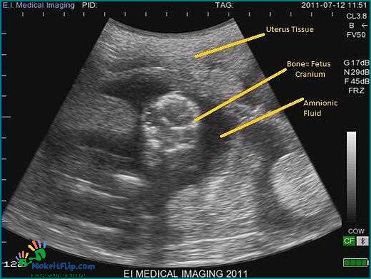
An ultrasound pic, short for ultrasound picture, is the visual representation of the ultrasound image. It provides a detailed view of the baby’s anatomy and can reveal important information about their growth and well-being. These images are often cherished by expectant parents as a way to bond with their unborn child and to share the excitement of the pregnancy with family and friends.
Interpreting an ultrasound pic requires specialized knowledge and expertise. Medical professionals, such as radiologists and obstetricians, are trained to analyze these images and identify any abnormalities or potential health concerns. They look for specific markers, such as the size and position of the baby, the presence of vital organs, and the flow of blood through the umbilical cord. This information helps them assess the baby’s overall health and make informed decisions about the pregnancy.
Understanding Ultrasound Pictures: A Comprehensive Guide

Ultrasound technology has revolutionized the field of diagnostic medical imaging, allowing healthcare professionals to obtain detailed images of the body’s internal structures. One of the most common applications of ultrasound is in pregnancy, where it is used to monitor the growth and development of the baby.
An ultrasound pic, also known as a sonogram, is a visual representation of the ultrasound image. It is created by sending high-frequency sound waves into the body and recording the echoes as they bounce back. These echoes are then transformed into a digital image that can be interpreted by medical professionals.
Understanding ultrasound pictures requires knowledge of the anatomy being imaged as well as the specific features and characteristics of the ultrasound technology being used. Medical professionals trained in ultrasound interpretation can identify and analyze various structures and abnormalities within the image.
During pregnancy, ultrasound pictures are commonly used to monitor the baby’s growth, check for any abnormalities or complications, and determine the baby’s position and sex. These pictures can provide valuable information to both the healthcare provider and the expectant parents.
It is important to note that ultrasound pictures are not just limited to pregnancy. They are also used in various other medical fields, such as cardiology, radiology, and obstetrics. In these areas, ultrasound can be used to visualize and diagnose conditions affecting the heart, liver, kidneys, and other organs.
When interpreting ultrasound pictures, medical professionals look for specific features and patterns that may indicate a particular condition or abnormality. They may use measurements, color coding, and other tools to assist in their analysis.
Overall, ultrasound technology has revolutionized the field of medical imaging, providing healthcare professionals with a non-invasive and safe way to visualize the body’s internal structures. Understanding ultrasound pictures requires specialized training and expertise, but they can provide valuable information for diagnosis and treatment planning.
What is an Ultrasound?

An ultrasound is a medical technology that uses sound waves to create images of the inside of the body. It is commonly used during pregnancy to monitor the development of the baby. The ultrasound scan, also known as an ultrasound pic, provides a visual representation of the baby and can be used to check for any abnormalities or potential issues.
During an ultrasound, a small handheld device called a transducer is used to emit high-frequency sound waves into the body. These sound waves bounce off the internal structures and create echoes, which are then captured by the transducer. The echoes are converted into an image that can be seen on a screen.
Ultrasound technology is safe and non-invasive, making it a preferred method for monitoring pregnancies. It allows healthcare professionals to visualize the baby’s growth, check the position of the placenta, and assess the overall health of the baby and the mother.
An ultrasound pic provides valuable information about the baby’s development and can be an exciting moment for expectant parents. It can show the baby’s size, shape, and movement, as well as reveal details such as the baby’s gender, if desired.
Overall, ultrasound technology plays a crucial role in prenatal care, allowing healthcare providers to monitor the progress of the pregnancy and ensure the well-being of both the mother and the baby.
Exploring the Basics of Ultrasound Imaging

Ultrasound imaging, also known as sonography, is a medical technology that uses high-frequency sound waves to create images of the inside of the body. It is commonly used in pregnancy to monitor the development of the baby and diagnose any potential issues.
During a pregnancy ultrasound, a handheld device called a transducer is used to send and receive sound waves. These waves bounce off the baby and other structures inside the mother’s body, creating an image that can be viewed on a screen.
The ultrasound image provides valuable information about the baby’s growth, position, and overall health. It can help doctors determine the baby’s age, estimate the due date, and detect any abnormalities or complications.
Ultrasound imaging is a non-invasive and safe diagnostic tool that does not use radiation, making it ideal for monitoring the progress of a pregnancy. It can be performed at different stages of pregnancy, with the first scan usually done around 8-12 weeks to confirm the pregnancy and establish the baby’s heartbeat.
As technology has advanced, ultrasound imaging has become more sophisticated, allowing for clearer and more detailed images. Three-dimensional (3D) and four-dimensional (4D) ultrasound scans provide a more realistic view of the baby’s features and movements.
In addition to pregnancy, ultrasound imaging is used in various medical fields to diagnose and monitor conditions affecting different parts of the body. It can be used to examine the abdomen, pelvis, heart, blood vessels, and other organs.
Overall, ultrasound imaging is a valuable tool in the field of medicine, providing doctors with important information for diagnosis and treatment. Its non-invasive nature and ability to produce real-time images make it an essential part of modern healthcare.
How Does Ultrasound Work?

Ultrasound is a diagnostic imaging technology that uses high-frequency sound waves to create images of the inside of the body. It is commonly used in medical settings, especially during pregnancy, to monitor the health and development of the fetus.
During an ultrasound scan, a small handheld device called a transducer is used to emit sound waves into the body. These sound waves bounce off the tissues and organs inside the body, creating echoes. The transducer then receives these echoes and converts them into electrical signals.
The electrical signals are then processed by a computer to create a visual representation of the internal structures. This image, known as an ultrasound pic or ultrasound image, can be viewed in real-time or saved for later analysis.
Ultrasound technology allows healthcare professionals to see the internal organs and tissues in a non-invasive way. It is particularly useful for examining soft tissues, such as the muscles, tendons, and organs in the abdomen and pelvis.
Ultrasound is also commonly used during pregnancy to monitor the growth and development of the fetus. It can provide valuable information about the baby’s position, size, and overall health. Additionally, ultrasound can be used to detect any abnormalities or potential complications.
In summary, ultrasound works by emitting high-frequency sound waves into the body and capturing the echoes that bounce back. These echoes are then converted into electrical signals and processed to create detailed images of the internal structures. This non-invasive and safe technology has revolutionized medical imaging and plays a crucial role in diagnosing and monitoring various conditions.
FAQ about topic Ultrasound Pic What You Need to Know and How to Interpret It
What is an ultrasound pic?
An ultrasound pic, or ultrasound image, is a picture that is created using ultrasound technology. It is a non-invasive medical procedure that uses sound waves to create images of the inside of the body.
How is an ultrasound pic taken?
An ultrasound pic is taken by a trained technician or doctor using an ultrasound machine. The machine emits high-frequency sound waves that bounce off the body’s tissues and organs, creating echoes. These echoes are then converted into images that can be seen on a screen.
What can an ultrasound pic show?
An ultrasound pic can show various things depending on the area of the body being examined. It can be used to visualize organs, such as the heart, liver, kidneys, and bladder. It can also be used to monitor the growth and development of a fetus during pregnancy.
How should I interpret an ultrasound pic?
Interpreting an ultrasound pic should be done by a trained healthcare professional, such as a radiologist or obstetrician. They will look for specific features and measurements to make a diagnosis or assess the health of the patient. It is important to consult with a medical professional for an accurate interpretation.
Are there any risks associated with getting an ultrasound pic?
Ultrasound imaging is generally considered safe and does not have any known risks. It does not use ionizing radiation like X-rays or CT scans. However, it is important to follow the guidelines and recommendations of your healthcare provider to ensure the safety and appropriateness of the procedure.
What is an ultrasound pic?
An ultrasound pic, also known as an ultrasound image or ultrasound scan, is a picture that is created using ultrasound technology. It is a non-invasive medical procedure that uses high-frequency sound waves to produce images of the inside of the body.
How is an ultrasound pic taken?
An ultrasound pic is taken by a trained healthcare professional using an ultrasound machine. The machine emits high-frequency sound waves, which are directed into the body using a handheld device called a transducer. The sound waves then bounce back off the internal organs and tissues, creating an image on a computer screen.
What can an ultrasound pic show?
An ultrasound pic can show various structures and organs inside the body, depending on the area being examined. It can be used to visualize the fetus during pregnancy, assess the health of organs such as the heart, liver, and kidneys, detect abnormalities or tumors, guide medical procedures, and monitor the progress of certain conditions.
How do you interpret an ultrasound pic?
Interpreting an ultrasound pic requires specialized training and expertise. A healthcare professional, such as a radiologist or obstetrician, will carefully analyze the image to identify and evaluate the structures or organs of interest. They will look for any abnormalities, measure certain dimensions, and compare the findings to normal ranges or previous images to make a diagnosis or determine the next course of action.
I’m Diana Ricciardi, the author behind Makeitflip.com. My blog is a dedicated space for mothers and their kids, where I share valuable insights, tips, and information to make parenting a bit easier and more enjoyable.
From finding the best booster seat high chair for your child, understanding the connection between sciatica and hip pain, to exploring the benefits of pooping in relieving acid reflux, I cover a range of topics that are essential for every parent.
My goal is to provide you with practical advice and solutions that you can easily incorporate into your daily life, ensuring that you and your child have the best possible experience during these precious years.
